MRI Odds and Ends...To Better Read a Scan
Picture this: You are in a MRI technologist program learning the foundation of what is needed to operate the MRI modality. It is time to discuss scanning.
How am I supposed to do it?
What do all the parameters mean?
There are how many pulse sequences?
What am I ACTUALLY looking at? (Sectional Anatomy, I’m looking at you…)
How do I figure out what level and plane I’m viewing?
Let’s just say the list can go on and on. There is of course a learning curve and it will become second nature as time goes on. This post compiles considerations for MRI scanning odds and ends to help put it all together to "read" a scan. What I mean by "reading" a scan is to understand what you are looking at to take the best possible images for the Radiologist doing the reading/interpreting. There is of course so much not included and still to be learned, but hopefully this blog makes the initial transition a little smoother.
Besides being able to operate the MRI correctly, we need to first know our anatomy.
Why?! How else would we know what we are looking at?
It looks a little different than the beautifully illustrated, perfectly proportional textbook images in COLOR. In the real world, humans do not have picture perfect anatomy and can have asymmetry (that is just their normal).
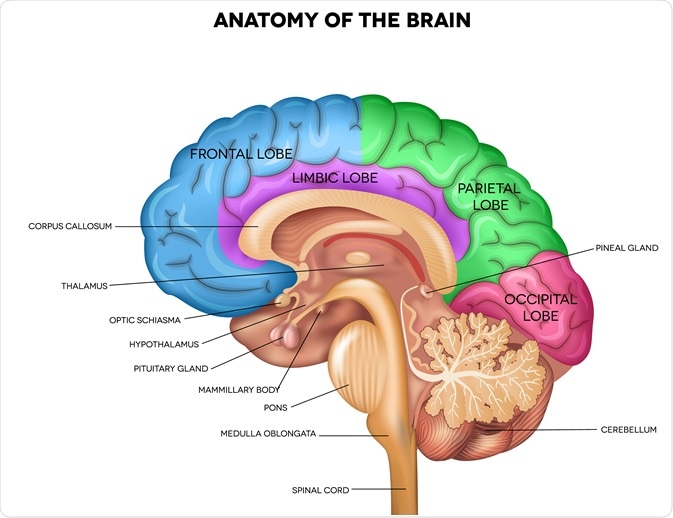 |
| Illustration from: Tefi/Shutterstock.com/Cuffari, 2020 |
Key take away: Each individual has their own unique characteristics and may make plotting for scans and viewing their images a little interesting. Maybe their frontal sinus is missing, or the lateral ventricles are asymmetrical (Bell, 2022) or oops, I counted three times and there seems to be only 4 lumbar vertebrae. Maybe there is a transitional vertebra and the L5 is fused to S1 or L1 is presenting as T12 (Konin & Walz, 2010).
Three planes…to see all the things from all the sides.
The body can be sliced and diced (hypothetically of course) into 3 anatomical planes:
CORONAL (aka Frontal)
SAGITTAL (aka Lateral)
AXIAL (aka Transverse)
 |
| 3 planes: Left-Axial, Middle- Coronal, Right- Sagittal Image from Research Gate |
You want to get all the view points and the best plotted images? Then these planes are needed. Can the anatomy look completely different; and/or weird, and you don't realize what you are looking at? ABSOLUTELY.
Besides viewing images and memorizing what they look like in different planes, there are a few more aspects that are beneficial.
Introducing our friend…the localizers. (Maybe sometimes referred to as scout or pilot images??)
Uh oh, are you lost in the anatomy or need a guide to figure out what level you are in the structure? Enter, the localizers! You and the localizers (party of four) need to work together as a group so you can see where you are in all three planes. This will make life easier in recognizing what the anatomy is and plotting.
Localizers are the initial images taken before running the various sequences in a protocol (Bruckner, 2016) so you:
A) Have the patient centered and correct anatomical landmarks
B) Have something to plot your images on because you can’t do it out of thin air
C) Have a reference point. As you scroll
through the slices, you can see the reference line move with you in whatever
plane you are in to see exactly where you are. As you run through your sequences, you can use the new scans instead of the localizers for plotting.
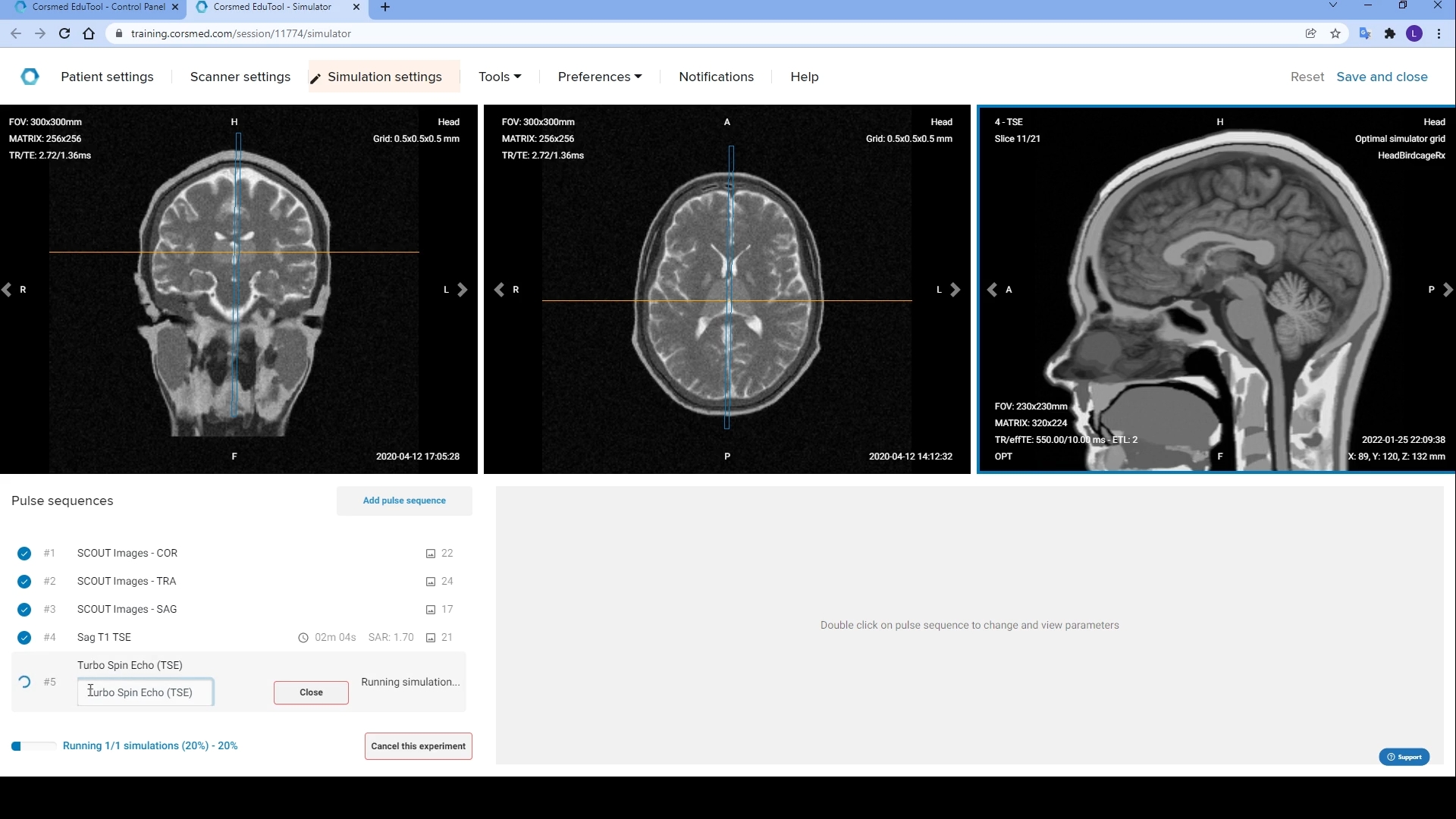 |
| MRI Simulator Localizers with blue and orange reference lines Image from Corsmed (Perneby, 2022) |
The images with reference points can help you identify the questionable slices from the money slices. To me, the “money” slice is when the anatomy is clear and you can tell what it is. It is the holy grail, “I know exactly what anatomical structure that is” slice.
The questionable/transitional slices to me are the ones where you are questioning whatever blob, dot or doodle you see and then find relief as you are scrolling and start to recognize it as you get towards the money slices.
Also, think about your context clues.
Is there any obvious give away?
What body part am I looking at?
Where is anterior/posterior, right vs left, superior vs inferior?
Once you are more comfortable with recognizing
anatomy, does anything look out of the ordinary?
I have my scan...Now what? Let's put it all together.
Below is a MRI sagittal brain that is labeled. Ideally, you would have all the slices to go through and identify vs a one slice wonder. You can scroll through the rest of the slices found on MRIMASTER.COM below:
MRIMASTER.COM MRI Sagittal Cross Sectional Anatomy of Brain
 |
| Sagittal MRI Brain labeled from MRIMASTER.COM |
I know, I know...there are quite a few structures labeled in the above image. Not only is it the money slice, but it does show us a lot of important anatomical structures in the brain.
Let's "read" this above slice:
- We are able to see and identify various anatomical structures in the brain including the corpus callosum, thalamus, midbrain, pons, medulla oblongata, pituitary gland, fourth ventricle, and so on (MRImaster.com).
- The image quality is clear with good contrast.
- We are able to see the whole head in the slice without cutting off any anatomy and the slice has coverage of the anatomical area (in this case the whole sagittal brain).
- We know where we are in the brain by knowing our sectional anatomy and can view our reference points (not pictured here) using the other planes just in case we forget.
- It will be ideally labeled, but we can see superior (top of head) and inferior (going into the neck) and anterior (front of face) and posterior (back of head) margins.
We did it!
You made it through all the sequences in the protocol with the correct anatomical coverage. Yay! Now, send it along, so the Radiologist can do their reading/interpretation.
This is not an all encompassing list. It is a relaxed and to the point interpretation of all the odds and ends needed to make "reading" a scan of an anatomical area (like the brain) a little smoother. This is just one of the steps in MRI to be able to take high quality images.
Happy Scanning!
References:
Bell, D. J. (2022, June 7). Asymmetry of the lateral ventricles. Radiopaedia Blog RSS. https://radiopaedia.org/articles/asymmetry-of-the-lateral-ventricles?lang=us
Bruckner, T. (2016, March 4). Sectional Anatomy for Radiographers.
Radiology Key. https://radiologykey.com/sectional-anatomy-for-radiographers/
Brain Illustration: Cuffari, Benedette. (2020, December 20). The Anatomy of the Human Brain. News-Medical. Retrieved on June 02, 2023 from https://www.news-medical.net/health/The-Anatomy-of-the-Human-Brain.aspx.
Konin, G. P., & Walz, D. M. (2010, November). Lumbosacral
transitional vertebrae: Classification, imaging findings, and clinical
relevance. AJNR. American journal of neuroradiology.
https://www.ncbi.nlm.nih.gov/pmc/articles/PMC7964015/
MRImaster Image: MRI Sagittal Cross Sectional Anatomy of Brain. MRImaster.com. (n.d.). https://mrimaster.com/anatomy%20brain%20sagittal.html
Corsmed Image: Perneby, A. (2022). MRI Brain with reference lines. MRI Brain
Protocol in Virtual MRI Simulator. Corsmed. Retrieved June 2, 2023, from
https://www.corsmed.com/2022/02/01/mri-brain-protocol-in-virtual-mri-simulator/.
3-planes Image: A Role of Medical Imaging Techniques in Human Brain Tumor Treatment - Scientific Figure on ResearchGate. Available from: https://www.researchgate.net/figure/MRI-planes-for-MRI-head-scan-a-Axial-b-Coronal-c-Sagittal-MR-scanner-can-generate_fig2_338448026 [accessed 1 Jun, 2023]
Comments
Post a Comment